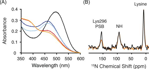
Figure 3.
Trapping of the Meta I intermediate in digitonin. (A) UV-Vis spectra provide a means to follow the conversion of rhodopsin (λmax = 500 nm, black line) to Meta I (λmax = 480 nm, blue line) after illumination by light (> 495 nm) at 4 °C. Meta I remains stable in digitonin for over 30 min at 4 °C (light blue line). Conversion at 20 °C leads to a mixture of Meta I and Meta II (red line), which does not change appreciably over 30 min (orange line). (B) One dimensional 15N spectra of rhodopsin (black) and Meta I (orange) labeled at 15Nζ-lysine are shown that were obtained using 1H-15N cross polarization. The 15Nζ-Lys2967.43 chemical shift is observed as a distinct narrow peak at 156.8 ppm in rhodopsin (black). In Meta I, the resonance shifts slightly and broadens. The 15Nζ resonances for the other 15Nζ-labeled lysines in rhodopsin are observed as a broad peak ~8.0 ppm.

