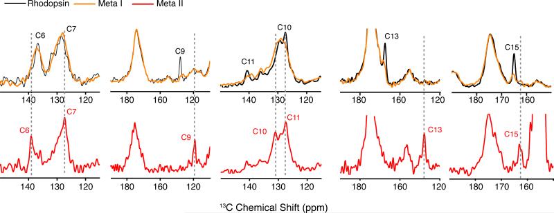
Figure 4.
One-dimensional 13C MAS NMR spectra of rhodopsin and Meta I. Rhodopsin was regenerated with 11-cis retinal selectively 13C labeled at different carbons along the polyene chain and the β-ionone ring. Overlap of the 13C MAS NMR spectra of rhodopsin (black) and Meta I (orange) shows that most of the retinal resonances broaden considerably in Meta I as compared to the sharp narrow resonances observed in rhodopsin and Meta II (red). There is nearly complete conversion of rhodopsin to Meta I. For example, the 13C9 retinal resonance in rhodopsin falls in an uncrowded region of the spectrum. The residual intensity of the 13C9 resonance in the Meta I spectrum is <10% of its original intensity in rhodopsin. Also, there is <10% conversion of rhodopsin to Meta II. For example, the spectra of Meta I containing 13C13-labeled retinal show a complete absence of a resonance associated with Meta II.

