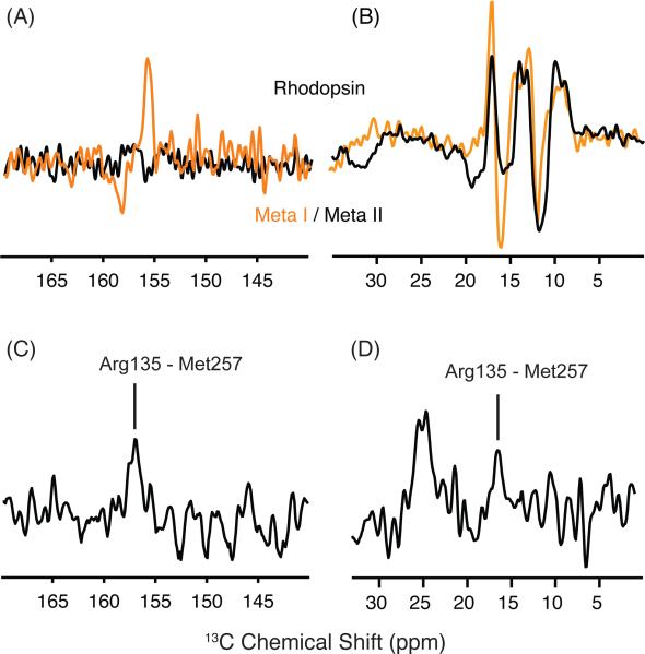
Figure 7.
13Cζ-arginine and 13Cε-methionine changes in the transition to Meta I. (A) Rhodopsin - Meta I (orange) and rhodopsin - Meta II (black) difference spectra are shown in the region of 13Cζ -arginine. The 13Cζ -arginine chemical shifts do not appear to differ between rhodopsin and Meta II, as indicated by the flat baseline in region of the difference spectrum. In contrast, the rhodopsin - Meta I difference spectrum reveals a single 13Cζ -arginine has changed in the transition to Meta I. (B) Rhodopsin - Meta I (orange) and rhodopsin - Meta II (black) difference spectra are shown in the region of 13Cε-methionine. A strong negative peak at 15.7 ppm corresponds to a new 13Cε-methionine resonance in Meta I. (C,D) Slices extracted from a 2D DARR NMR spectrum reveal a cross peak between 13Cζ -Arg1353.50 and 13Cε-Met2576.40 in Meta I on both sides of the diagonal in 2D spectrum.

