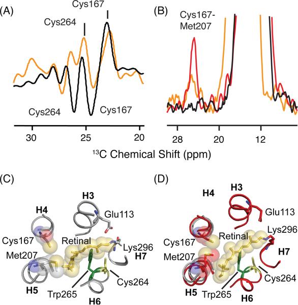
Figure 9.
Chemical shift changes in 13Cβ-cysteine in the transition from rhodopsin to Meta I. (A) Rhodopsin - Meta I (orange) and rhodopsin - Meta II (black) difference spectra in the region of reduced 13Cβ-cysteine resonances. (B) Slices taken through 2D 13C DARR NMR spectra of rhodopsin (black), Meta I (orange) and Meta II (red) exhibit a cross-peak between Met2075.42 and Cys1674.56. The intensity of the Cys1674.56 - Met2075.42 cross-peak in Meta I is intermediate between rhodopsin and Meta II, suggesting that there is partial rotation of H5. (C) Structure of rhodopsin (PDB access code = 1GZM) in the region of the retinal binding site. The structure highlights the positions of Cys1674.56 and Met2075.42. (D) Structure of Meta II (PDB access code = 3PXO) showing displacement of H5 and closer interaction of Cys1674.56 and Met2075.42.

