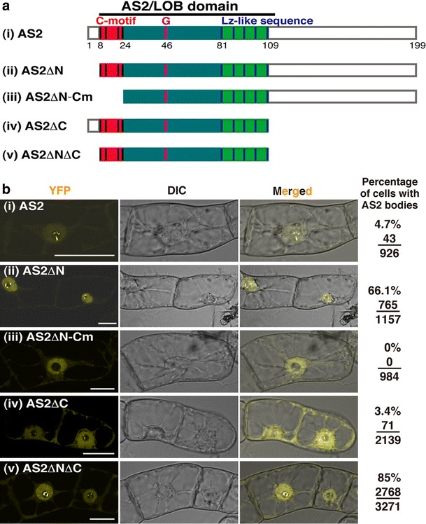
Fig. 2.

Subcellular localization of wild-type AS2 and its deletion mutants that were fused to YFP. a Schematic representation of wild-type and deletion mutants. Predicted domain organization and relevant amino-acid positions for AS2 are indicated above and below, respectively, in the wild-type schematics. Coding sequences for all AS2 proteins (i–v) were fused to the sequence corresponding to the N-terminus of the YFP sequence. These fusion constructs were linked to the estrogen-inducible promoter. b Subcellular localization of deletion mutants of AS2 in interphase cells of transformed BY-2 lines. The transformed cells harboring these fusion constructs were incubated for 16 h in the presence of 0.05 μM 17-β-estradiol. Living cells were observed by confocal fluorescence microscopy to detect fluorescence of YFP (yellow YFP). Nomarski (DIC) and merged images (Merged) are also shown. Numbers on the right represent ratios of cells showing AS2 bodies to interphase cells examined. Bars 20 μm
