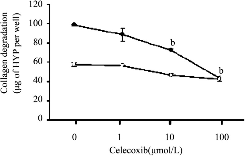
Figure 1. Dose-dependent inhibition effect by Celecoxib of IL-1ß-induced collagen degradation by corneal fibroblasts.
Rabbit fibroblasts were cultured in collagen gels in the presence of plasminogen, in the absence (open symbols)) or presence (closed symbols) of IL-1ß (0.1ng/mL) and in the presence of the indicated concentrations of Celecoxib. After incubation of the cells for 48 hours, the amount of degraded collagen in the culture supernatants was determined. Data are expressed as micrograms of hydroxyproline (HYP) per well and are means±SEM of values from an experiment that was repeated a total of three times with similar results. bP< 0.001 (Dunnett's test) vs the value for cells cultured with IL-1ß in the absence of Celecoxib.

