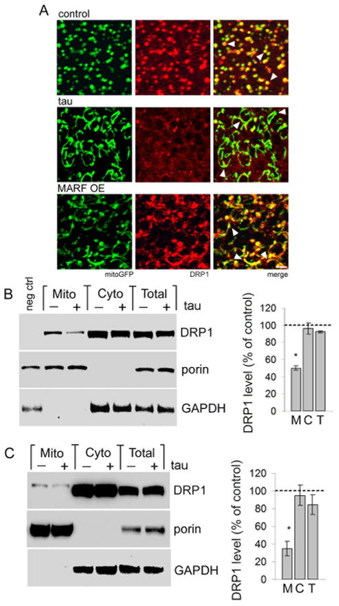
Figure 3. Tau expression blocks mitochondrial localization of DRP1.
(A) Mitochondria (mitoGFP, green) and HA-tagged DRP1, detected with an antibody to HA (red), colocalize in control neurons (control, arrowheads). Mitochondria in tau transgenic neurons show markedly reduced colocalization with DRP1 (tau, arrowheads). Elongated mitochondria produced by overexpression of MARF retain the ability to recruit DRP1 (MARF OE, arrowheads). Scale bar is 2 μm. Control is elav-GAL4/+; UAS-mitoGFP/+; HA-DRP1/+. (B) Subcellular fractionation of fly head homogenates shows depletion of HA-DRP1, detected with an antibody to HA, from the mitochondrial fraction of tau flies (Mito, M) with equivalent levels of DRP1 in the cytoplasmic fraction (Cyto, C) or total homogenate (Total, T). Control is elav-GAL4/+; HA-DRP1/+, negative control (neg ctrl) is elav-GAL4/+. (C) Subcellular fractionation of mouse hippocampal homogenates shows reduction of mitochondrially-localized DRP1 in tau animals (Mito, M), while total (Total, T) and cytoplasmic (Cyto, C) DRP1 levels are not significantly affected. The blots in (B,C) are reprobed for porin and GAPDH to demonstrate enrichment of mitochondrial and cytoplasmic proteins in the appropriate fractions. The graphs in (B,C) show DRP1 levels as percent of control in each fraction from tau transgenic flies or mice. Asterisks indicate P < 0.01, one-way ANOVA with Student-Neuman-Keuls test, n = 3. See also Fig. S3.

