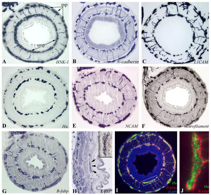
Figure 6. Colorectal ENS at E12-E14 (HH38–40).
Immunohistochemistry was performed on transverse sections of E14 (HH40) mid-colorectum with HNK-1 (A), N-cadherin (B) and L1CAM (C). Differentiated ENCCs express neuronal markers Hu (D), NCAM (E), and neurofilament (F), and glial markers B-fabp (G) and GFAP (H). The submucosal ganglion marked with arrows is magnified in the inset (H). The close relationship between neurons (Hu) and glia (B-fabp) is seen at E12 (HH38; I, box magnified in J).

