
Figure 1.
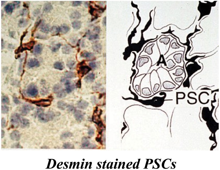
Pancreatic stellate cells in rat pancreas stained for the selective marker desmin. The left panel shows a representative photomicrograph of normal rat pancreas immunostained for desmin. The right panel depicts the corresponding line diagram. Desmin positive PSCs with long cytoplasmic projections are located at the basolateral aspect of acinar cells (A). Reprinted with permission from BMJ Group.