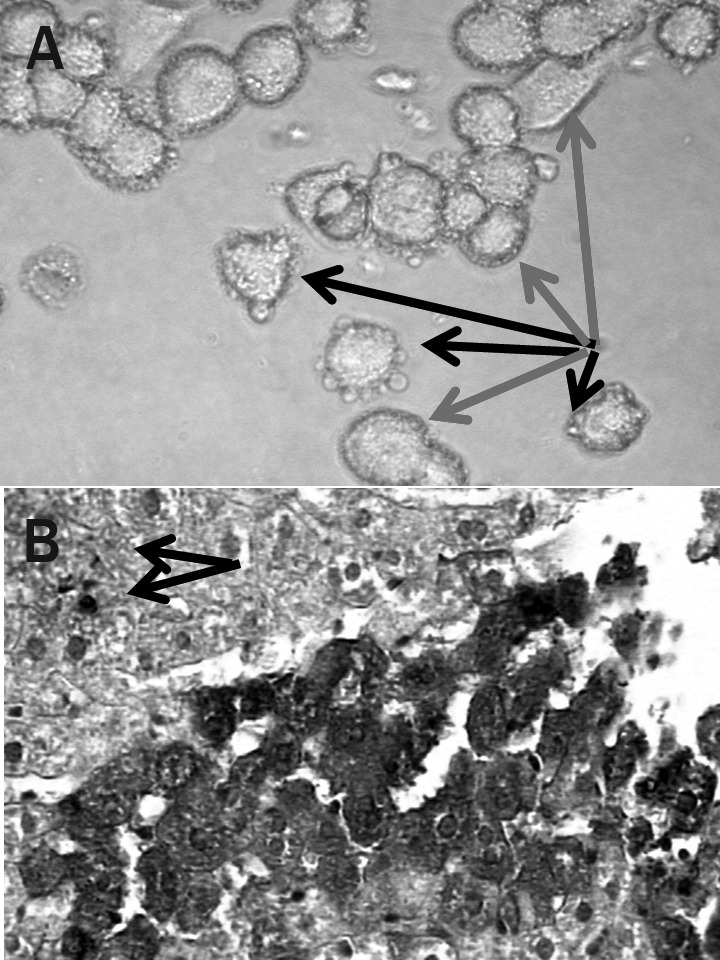
Figure 2.

Microphotographs revealing individuality of the cellular 4-hydroxy-2-nonenal (HNE). Although standardized line HeLa cells (A) are considered all to be identical, a treatment with 50 μM HNE for 30 minutes revealed great differences in their individual reactivity to the toxic dose of the aldehyde. Thus, while some cells showed membrane lipid peroxidation damage (“blebs” indicated by black arrows), their neighbor cells did not show any signs of the damage (gray arrows). Similarly, 30 minutes after biopsy needle puncture the affected liver cells (B) showed strong immunopositivity for HNE (darker shade), while their neighbor cells were negative for the aldehyde (lighter shade). However, some cells, even remote, began generating HNE indicating individual sensitivity of the cells to the damage signaling (indicated by the black arrows).
