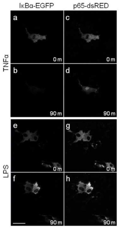
Figure 4. Nuclear translocation of p65 or IκBα degradation in granulosa cells was induced by TNFα but not LPS.
Granulosa cells were cultured in glass bottom petri dishes and transfected with IκBα-EGFP (a-b, e-f) and p65-dsRed (c-d, e-h). Live cell confocal microscopy under culture conditions was performed to track degradation of IκBα-EGFP and nuclear translocation of p65-dsRed. Cells were treated with 20 ng/ml TNFα (a-d) as a positive control, or 1 μg/ml ultrapure LPS (e-h). Scale bar represents 50μm.

