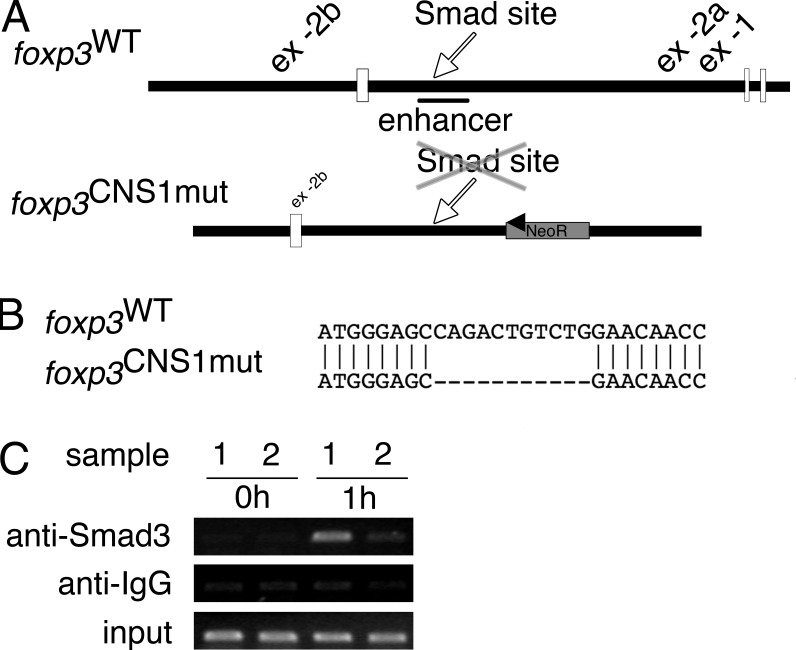
Figure 1.
Deletion of Smad-binding site in foxp3 enhancer/CNS1. (A) Schematic depiction of upstream region of wild-type foxp3 locus and the gene targeting vector. The CNS1 and the position of the Smad-binding site in the wild type or deleted binding site in the foxp3CNS1mut allele are indicated. (B) Alignment of the wild type and the foxp3CNS1mut allele at the position of the Smad-binding site, showing the deleted sequence. (C) ChIP analysis of Smad3 binding to the foxp3 CNS1 region. foxp3WT (sample 1) or foxp3CNS1mut (sample 2) CD4+CD25− splenocytes were not stimulated (0 h) or stimulated for 1 h with TGF-β, anti-CD3, and anti-CD28. Immunoprecipitated DNA was analyzed by PCR. Data are representative of two independent experiments.

