Figure 8.
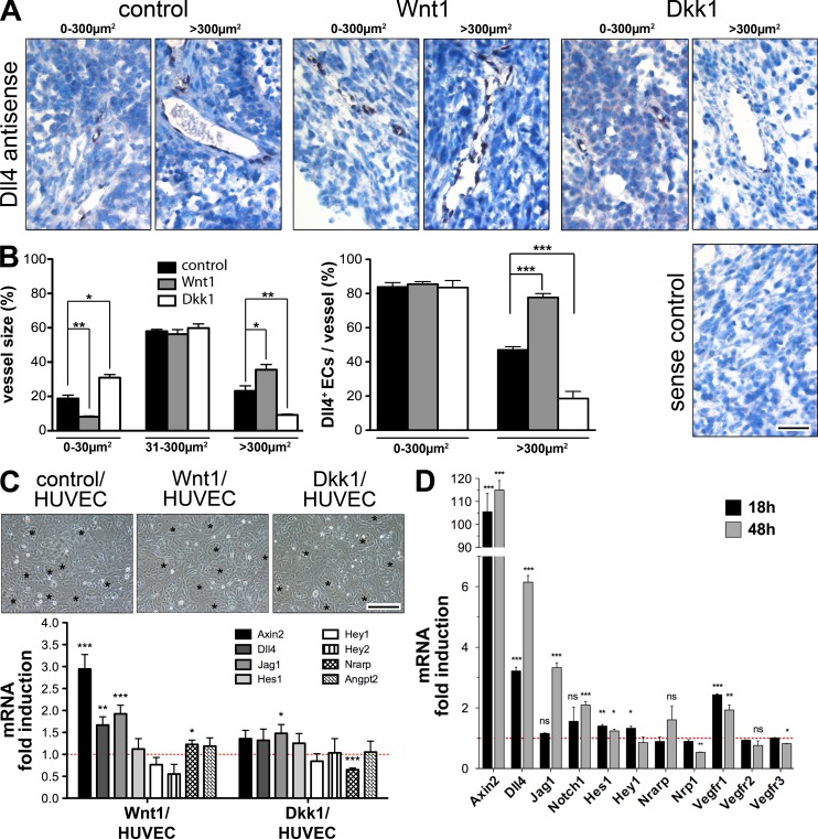
Glioma-derived Wnt1 up-regulated endothelial Dll4, leading to a stalk cell–like gene signature. (A) ISH against Dll4 (black) and hematoxylin (blue) of subcutaneous control−D, Wnt1−D, and Dkk1−D GL261 tumors, showing representative small (0–300 µm2; left) and large (>300 µm2; right) vessels. (B, left) Quantification of vessel diameter (n = 3 tumors/group, 15 pictures/tumor) grouped in categories of 0–30 µm2, 31–300 µm2, and >300 µm2 (*, P < 0.05; **, P < 0.01). (right) Quantification of Dll4+ ECs (n = 6 tumors/group, 5 small and 5 large vessels/tumor; ***, P < 0.001). (C, top) HUVECs co-cultivated with control−D, Wnt1−D, and Dkk1−D GL261 cells (asterisks indicate HUVECs). (bottom) qRT-PCR (n = 6) for human genes regulated by Wnt1−D and Dkk1−D compared with control−D co-cultures (red line). (D) qRT-PCR for mouse genes regulated in MBEs stimulated by Wnt3aCM for 18 and 48 h compared with controlCM (red line; n = 3; *, P < 0.05; **, P < 0.01; ***, P < 0.001). Bars: (A) 50 µm; (C) 80 µm. Error bars indicate SEM.

