Figure 8.
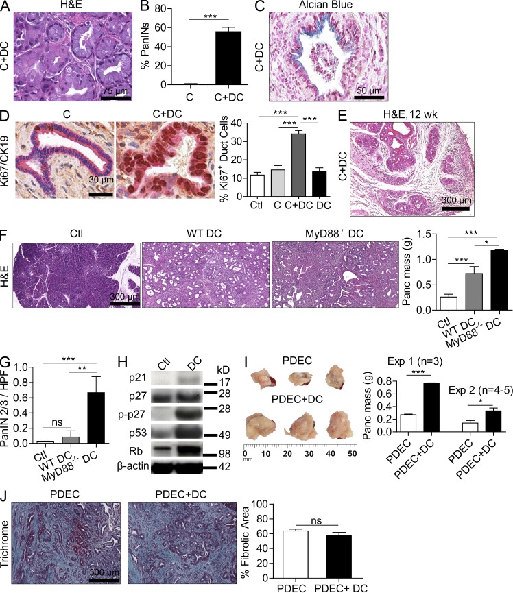
DCs promote the transition from chronic pancreatitis to ductal dysplasia and accelerate the growth rate of pancreatic tumors. (A and B) 4-wk-old WT mice were treated with C or C+DCs for 3 wk. Representative H&E-stained section from C+DC-treated pancreata is shown. The fraction of pancreatic ducts in C or C+DC mice exhibiting PanIN lesions was quantified by examining pancreata from >15 mice per group. (C) Representative paraffin-embedded section of pancreata from mice treated with C+DCs and stained with Alcian blue is shown. (D) Pancreata from mice treated with C or C+DCs were stained for CK19 (pink) and Ki67 (brown), and the fraction of Ki67+ ductal cells was quantified for each treatment group. (E) Representative H&E-stained image of pancreas from a mouse treated for 12 wk with C+DCs. (F and G) 4-wk-old p48Cre;KrasG12D mice were adoptively transferred with DCs derived from WT or MyD88−/− mice thrice weekly for 5 wk before sacrifice (n = 5/group). Representative H&E-stained sections are shown, pancreata were weighed, and the fraction of PanIN 2/3 ducts per HPF was determined (G). (H) Lysate from pancreata of mice adoptively transferred with WT DCs or saline was tested for expression of p21, p27, p-p27, p53, and Rb by Western blotting. (I) Mice were challenged with 3 × 10⁶ (experiment 1) or 106 (experiment 2) KRasG12D PDECs. In each experiment, selected mice were adoptively transferred with DCs (106) thrice weekly from weeks 2–6. Mice were sacrificed after 6 wk, and pancreatic lesions were weighed. (J) Representative Trichrome-stained paraffin-embedded sections are shown, and the fraction of fibrotic pancreatic area was quantified (*, P < 0.05; **, P < 0.01; ***, P < 0.001). Error bars indicate standard error of the mean.

