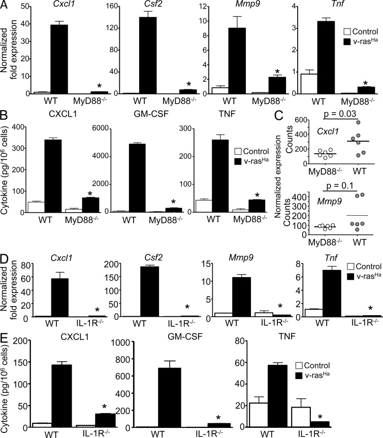
Figure 2.
Expression of NF-κB–regulated proinflammatory factors in RAS-transformed keratinocytes requires a functional IL-1–MyD88 axis. (A and D) Real-time PCR analyses 3 d after RAS transduction of Cxcl1, Csf2, Mmp9, and Tnf mRNA expression in control and v-rasHa–transduced keratinocytes (WT and MyD88−/− [A] or IL-1R−/− [D]). (B and E) CXCL1, GM-CSF, and TNF concentrations were determined by ELISA in culture supernatants from control and v-rasHa–transduced keratinocytes (WT and MyD88−/− [B] or IL-1R−/− [E]) 3 d after RAS transduction. Data shown are representative of three independent experiments, and bars represent the mean ± SEM of five replicates. *, P < 0.05 between v-rasHa WT and v-rasHa gene–deficient strain. (C) NanoString analysis of Cxcl1 and Mmp9 mRNA expression in laser capture–microdissected WT and MyD88−/− papillomas from chemically induced skin carcinogenesis. The y axis shows normalized NanoString counts. Each circle represents an independent squamous papilloma. Horizontal bars indicate the mean.

