Figure 4.
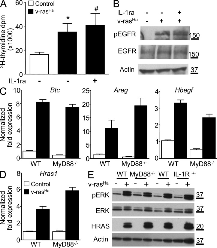
The EGFR autocrine loop is intact in RAS-transformed MyD88-deficient keratinocytes. (A) Tritiated thymidine incorporation was measured in control and v-rasHa–transduced WT keratinocyte cultures treated with IL-1ra for 3 d. Data shown are representative of three independent experiments, and bars represent the mean ± SEM value of four replicates. *, P < 0.05 between v-rasHa and control; #, no significant difference between v-rasHa − IL-1ra and v-rasHa + IL-1ra. (B) Total cell extract from primary keratinocytes transduced for 3 d with v-rasHa in the presence or absence of IL-1ra (Anakinra) was analyzed by Western blotting for phospho-EGFR (Tyr1068), total EGFR, and actin as loading control. The picture is representative of three independent experiments. (C) Real-time PCR analysis of Btc (betacellulin), Areg (amphiregulin), and Hbegf (heparin-binding EGF-like growth factor) in control and v-rasHa–transduced keratinocytes (WT and MyD88−/−) 3 d after RAS transduction. (D) Real-time PCR analysis of Hras1 (H-ras) in control and v-rasHa–transduced WT and MyD88−/− keratinocytes. (C and D) Data shown are representative of three independent experiments, and bars represent the mean ± SEM of three replicates. (E) Total cell extract from MyD88−/− and IL-1R−/− primary keratinocytes and their respective controls transduced for 3 d with v-rasHa were analyzed by Western blotting for phospho-ERK, total ERK, H-ras, and actin as loading control. The picture is representative of two independent experiments. (B and E) Molecular mass is indicated in kilodaltons.

