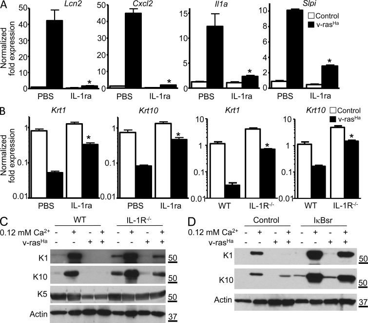
Figure 5.
IL-1α–mediated activation of NF-κB is responsible for the altered expression of differentiation markers in RAS-transformed keratinocytes. (A) Real-time PCR analysis of Lcn2, Cxcl2, Il1a, and Slpi mRNA expression in control or v-rasHa–transduced WT keratinocytes treated with PBS or IL-1ra (Anakinra). *, P < 0.05 between v-rasHa PBS and v-rasHa IL-1ra. (B) Real-time PCR analysis of Krt1 (keratin 1) and Krt10 (keratin 10) mRNA expression in control and v-rasHa–transduced keratinocytes (PBS or IL-1ra treated or IL-1R WT and IL-1R−/−) 3 d after RAS transduction. *, P < 0.05 between v-rasHa PBS and v-rasHa IL-1ra or v-rasHa IL-1R WT and v-rasHa IL-1R−/−. (A and B) Data shown are representative of three independent experiments, and bars represent the mean ± SEM of three replicates. (C and D) Total SDS cell extracts from control and v-rasHa–transduced keratinocytes (IL-1R WT and IL-1R−/−; C) or WT keratinocytes infected with A-CMV (control) or degradation-resistant IκBα super repressor (IκBsr) adenovirus to block NF-κB activity (D) were analyzed through immunoblotting with specific antibodies for the expression of basal (K5) and early markers of differentiation (K1 and K10). SDS lysates were analyzed from 3-d posttransduction cultures that were maintained in 0.05 or 0.12 mM Ca2+ media for an extra 24 h. Data are representative of three independent experiments. Molecular mass is indicated in kilodaltons.

