Figure 6.
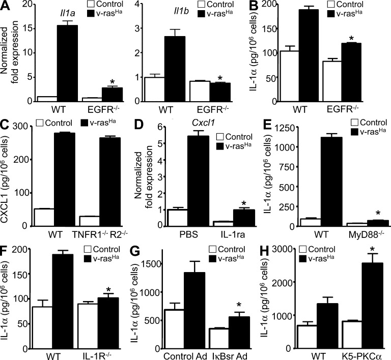
Oncogenic RAS-mediated EGFR–IL-1R signaling loops. (A) Real-time PCR analysis of IL-1α (Il1a) and IL-1β (Il1b) mRNA expression in control or v-rasHa–transduced WT and EGFR−/− keratinocytes. (B) Culture supernatants from WT or EGFR−/− primary keratinocytes were collected after control or v-rasHa transduction. IL-1α concentrations were determined by ELISA. *, P < 0.05 between v-rasHa EGFR WT and v-rasHa EGFR−/−. (C) Culture supernatants from WT and TNFR1−/−/TNFR2−/− primary keratinocytes were collected after control or v-rasHa transduction. CXCL1 concentrations were determined by ELISA. (D) Real-time PCR analysis of CXCL1 (Cxcl1) mRNA expression in keratinocytes pretreated with PBS or IL-1ra (Anakinra) for 1 h before TGF-α stimulation for an extra hour. *, P < 0.05 between TGF-α treatment − IL-1ra and TGF-α treatment + IL-1ra. (E and F) Culture supernatants from WT or MyD88−/− primary keratinocytes were collected after control or v-rasHa transduction. IL-1α concentrations were determined by ELISA in culture supernatants from control and v-rasHa–transduced keratinocytes (WT and MyD88−/− [E] or IL-1R−/− [F]) 3 d after v-rasHa transduction. *, P < 0.05 between v-rasHa WT and v-rasHa gene–deficient strain. (G) Culture supernatant collected from control or v-rasHa–transduced keratinocytes infected with A-CMV (control Ad) or degradation-resistant IκBα (IκBsr Ad) adenovirus to block NF-κB activity. IL-1α concentrations were determined by ELISA. *, P < 0.05 between v-rasHa control Ad and v-rasHa IκBsr Ad. (H) Culture supernatants from WT or primary keratinocytes from mice overexpressing PKC-α (K5-PKCα) were collected after control or v-rasHa transduction. IL-1α concentrations were determined by ELISA. *, P < 0.05 between v-rasHa WT and v-rasHa K5-PKCα. Data shown are representative of two (C and H) to three (A, B, and D–G) independent experiments, and bars represent the mean ± SEM of three (A, B, and D–H) to five (C) replicates.

