
Figure 5.
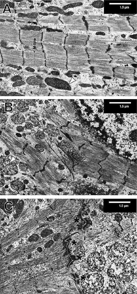
SNP alters the ultra-structure of cardiomyocytes. Isolated ARVM were cultured for 14 days then treated with 3 mM SNP for 0 (A), 3 (B) and 5 (C) hours and processed for electron microscopy. Typical cells are shown out of 15 areas for each condition from 3 independently processed hearts. Mitochondrial swelling was observed in SNP-treated cultures. Typical mitochondria are indicated with black arrowheads (A–C). Fasciae adherens and desmosomes are visible in the middle of image C (black asterisks) indicating a line of cell-cell contacts between two cardiomyocytes differently affected by the SNP treatment.