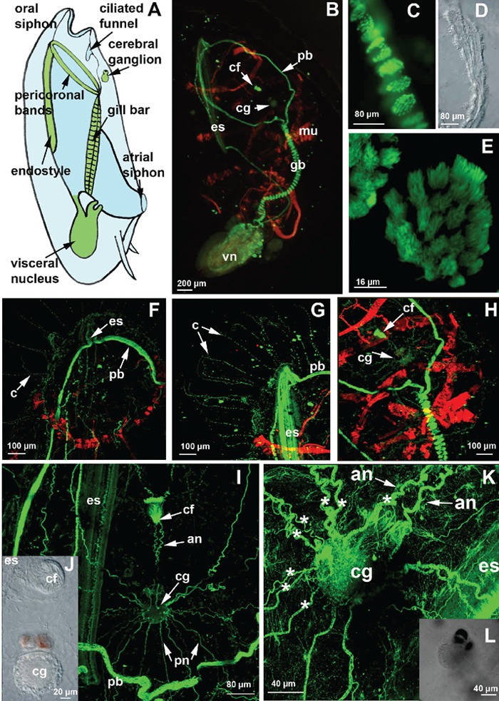
Figure 2.

Whole-mount labelling by FITC-conjugated anti-tubulin antibodies and by TRITC-conjugated phalloidin. A) schematic drawing of the lateral view of a blastozooid (redrawn after 4); B) whole-mount specimen observed using a fluorescent microscope; C,E) confocal laser microscope (CLM) images of the ciliary apparatus of the gill bar at different magnifications, which were obtained by the sum of 30 and 20 optical sections, step size of 1 µm. D) transmission microscope image of the gill bar; F,G) CLM images of the anterior end of a specimen immunolabelled with an anti-acetylated tubulin antibody, showing the long cilia passing through the tunic; H,I,K) CLM images at different magnifications of the cerebral ganglion labelled with anti-β-tubulin antibody; K) CLM image of the cerebral ganglion in which the 8 main fibres of the left side are indicated by asterisks; J) details using light microscopy with Normasky optics of the cerebral ganglion and of the ciliated funnel; L) transmission microscope image of the samples observed in K at a lower magnification. an, anterior nerves; cf, ciliated funnel; cg, cerebral ganglion; c, cilia; cf, ciliated funnel; es, endostyle; gb, gill bar; mu, muscle band; pb, pericoronal band; pn, posterior nerves; vn, visceral nucleus.
