
Figure 10.
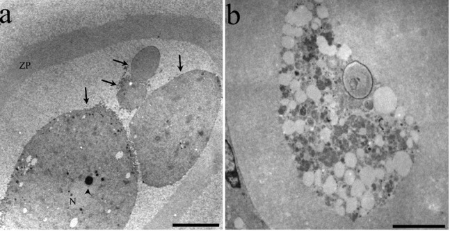
Ultrastructural features of atretic oocytes in antral follicles: a) a highly altered oocyte in the apoptotic process; the oocyte is fragmented (arrows) one of the cellular fragments has a substantially altered nucleus (N) with a highly compacted nucleolus (arrowhead). The zona pellucida (ZP) is completely detached from the oocyte; b) oocyte in the autophagic process of cell death, evidenced by numerous autophagic vesicles in different degrees of degradation. Uranyl acetate and lead citrate. Scale bars: a, 10 microns; b, 5 µm.