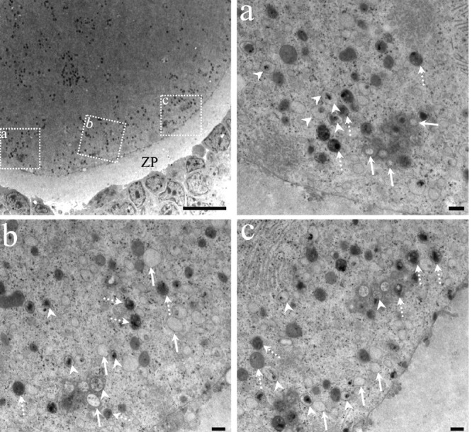
Figure 11.

Ultrastructural features of an oocyte in the autophagic cell death process. The relationship between the granulosa cells and the oocyte are highly altered; there are no cytoplasmic prolongations of the granulosa or microvillus of the oocyte. The zona pellucida (ZP) surrounds a highly altered oocyte with numerous dense structures. The dotted squares have been enlarged in micrographs a, b and c. High magnification pictures of autophagic vesicles in different degrees of degradation of their content. Arrows point to clear autophagic vesicles containing highly degraded material; arrowheads point to autophagic vesicles containing debris in an intermediate degree of degradation. The autophagic vesicles with recently sequestered material are indicated by dotted arrows. Scale bars: low magnification 10 microns; a, b, and c, 500 nm.
