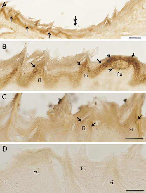
Figure 3.

Double immunoperoxidase staining of TRPV1 and CGRP in the rat tongue. A) Clear TRPV1 immunoreactive (ir) epithelium (arrows) are observed in apex region (left side of the panel A) of the tongue; note pale immunoreactivity of the TRPV1 in posterior region (right side of the panel A), double arrows indicate an approximate intersectional region; the double arrows correspond to double arrows in the Figure 4 A. B) Higher magnification of apex region; arrows showing TRPV1-ir epithelium; arrowheads showing CGRP-ir terminals, small black dot like structure, around TRPV1-ir epithelium of the fungiform papilla (see also Figures 4 and 5); C) higher magnification of posterior region; weakly TRPV1-ir is observed (arrows); D) control section by omitting the primary antibodies. Fi, filiform papilla; Fu, fungiform papilla. Scale bars: A) 100 µm; B–D) 50 µm.
