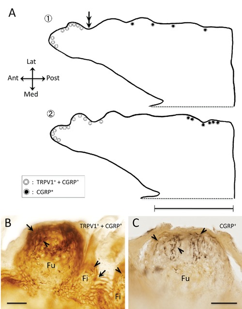
Figure 4.

Distributions of TRPV1 immunoreactive (ir) and CGRP-ir in the fungiform papilla in the rat tongue. A) Line drawings of sagittal section of the tongue; the two line drawings are reconstructed from total 71 sections, the section no. 1 consists 35 sections and no. 2 consists 36 sections respectively, and depicted approximately 2 mm extent from midline to lateral; white and black circles indicating the fungiform papilla distribution on the tongue; white circles, strongly TRPV1-ir epithelium (arrow in panel B) and CGRP-ir terminals (arrowheads in panel B) distributing around the epithelium; black circles, CGRP-ir terminals (arrowheads in panel C) distributing around the epithelium, which is not TRPV1-ir epithelium (C); note concentration of white circles in apex region; double arrows indicate an approximate posterior border of the concentration and correspond to double arrows indicating in the Figure 3A; B,C) photographs of the two cases of fungiform papilla, B corresponding to the white circle and C corresponding to the black circle respectively. Ant, anterior; Fi, filiform papilla; Fu, fungiform papilla; Lat, lateral; Med, medial; Post, posterior. Scale bars: A) 5 mm; B,C) 30 µm.
