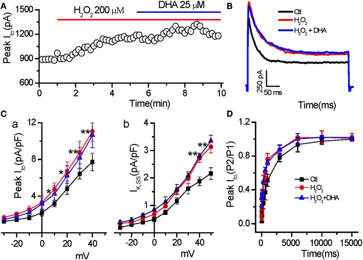
Figure 4.
Less effect of DHA on Ito enhanced by H2O2. (A) Time course of peak Ito in a myocyte treated with H2O2 in the absence and presence of DHA. (B) Representative traces of the Ito under control, in the presence of H2O2 (200 μM), and H2O2 + DHA (25 μM), respectively. (C) Current–voltage relations of the peak Ito (C-a) and steady state currents (IK,SS, C-b) showing less effects of DHA on enhancement of peak Ito and IK.ss (n = 6, *p < 0.05, **p < 0.01 vs. control.). Test potentials ranged from −60 to +50 mV in 10 mV steps. (D) Recovery of Ito from inactivation showing no significant effect of DHA (25 μM) on the Ito recovery sped-up by H2O2 (200 μM; p > 0.05, n = 7).

