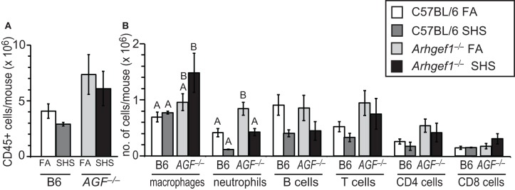
Figure 2.
Leukocyte numbers in lung tissue after 4 weeks of second hand smoke exposure. (A) Total number of CD45+ leukocytes in enzymatically digested and lavaged lung tissue from C57BL/6 (B6) and Arhgef1−/− (AGF−/−) mice exposed to filtered air (FA) or second hand smoke (SHS) for 4 weeks. (B) Number of leukocytes within different populations from enzymatically digested and lavaged lung tissue. Shown are the number of macrophages (F4/80+), neutrophils (Gr-1+), B lymphocytes (B220+) and T lymphocytes (CD3+), including CD4+ and CD8+ cells. Three month old C57BL/6 (B6) mice exposed to filtered air (FA) (open bars, n = 8), 3 month old C57BL/6 (B6) mice exposed to SHS for 4 weeks prior to harvest (dark gray bars, n = 4), 3 month old Arhgef1−/− (AGF−/−) mice exposed to filtered air (FA) (light gray bars, n = 7) and 3 month old Arhgef1−/− (AGF−/−) mice exposed to SHS for 4 weeks prior to harvest (black bars, n = 4). Data represents mean ± SE. A One-Way ANOVA was performed on leukocyte populations. Statistically significant differences between groups were detected only for macrophages and neutrophils. A post hoc Tukey–Kramer HSD t test was performed on these groups. Groups not sharing the same letter are significantly different, P < 0.05. For the macrophages the B6 FA, B6 SHS, and AGF−/− FA groups all share the letter A so none of these groups are significantly different from each other. The AGF−/− FA and the AGF−/− SHS groups share the letter B so these two groups are not significantly different from each other. The significant difference in macrophages occurs between the AGF−/− SHS group (black bar) which only has the letter B designation and the B6 FA and B6 SHS groups (open and light gray bars) which only have the letter A designation. Cells were enumerated and analyzed as described in materials and methods.

