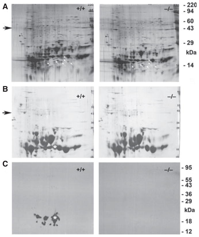
Fig. 3.

Two-dimensional gel electrophoresis of WT and KO mouse lenses. (A) Silver-stained blots of WT and KO lenses. White arrows indicate the positions of spots missing in the KO lens. The black arrow indicates the size region for actin. (B) Coomassie stain and (C) western blot of γS for duplicate 2D gels of WT and KO lenses. The asterisk shows the position of the main γS spot. The WT lens shows evidence of multiple post-translational modifications of γS, whereas the KO lens shows complete absence of γS immunoreactivity.
