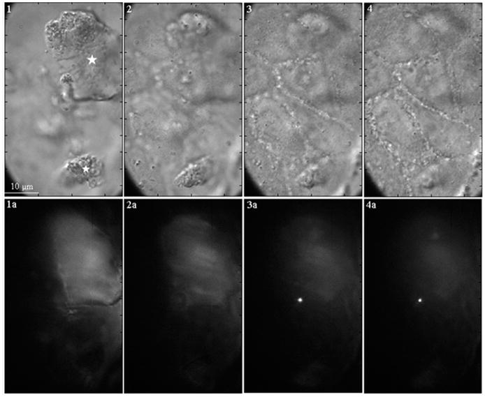
FIGURE 3.
DIC image (1) showing the presence of two large dye-tagged SAMMS particles (about 10 and 25 μm, indicated by the stars) on the cell surface. As images are taken deeper in the cells (2–4), the particles disappear. The correlated fluorescence image (1a and 2a) shows dim or no fluorescence signals from the dye-tagged particles as a result of fluorescence quenching by Trypan Blue. Together, these observations indicate that the large particles stay at the cell surface. The fluorescence images that are taken deeper in the cells (3a and 4a) show the presence of a bright spot, which indicates that a small particle (~1–2 μm) was internalized into the cytoplasm, where it was protected from the quenching by Trypan Blue.

