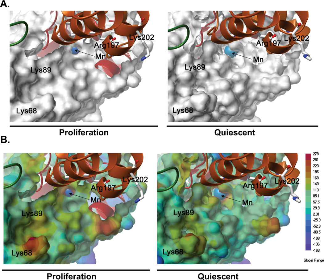
Figure 6. Structural and electrostatic changes in MnSOD during quiescence and proliferation.
(A) Connolly molecular surface representations of a monomeric unit of the methylated-MnSOD tetramer in proliferation and quiescence; the manganese (Mn) ion (blue) is shown as a space fill representation. (B) Electrostatic potential energy surfaces of the methylated-MnSOD tetramer in proliferation and quiescence; surfaces colored blue are negative and the Mn ion (blue) is shown as a space fill representation.

