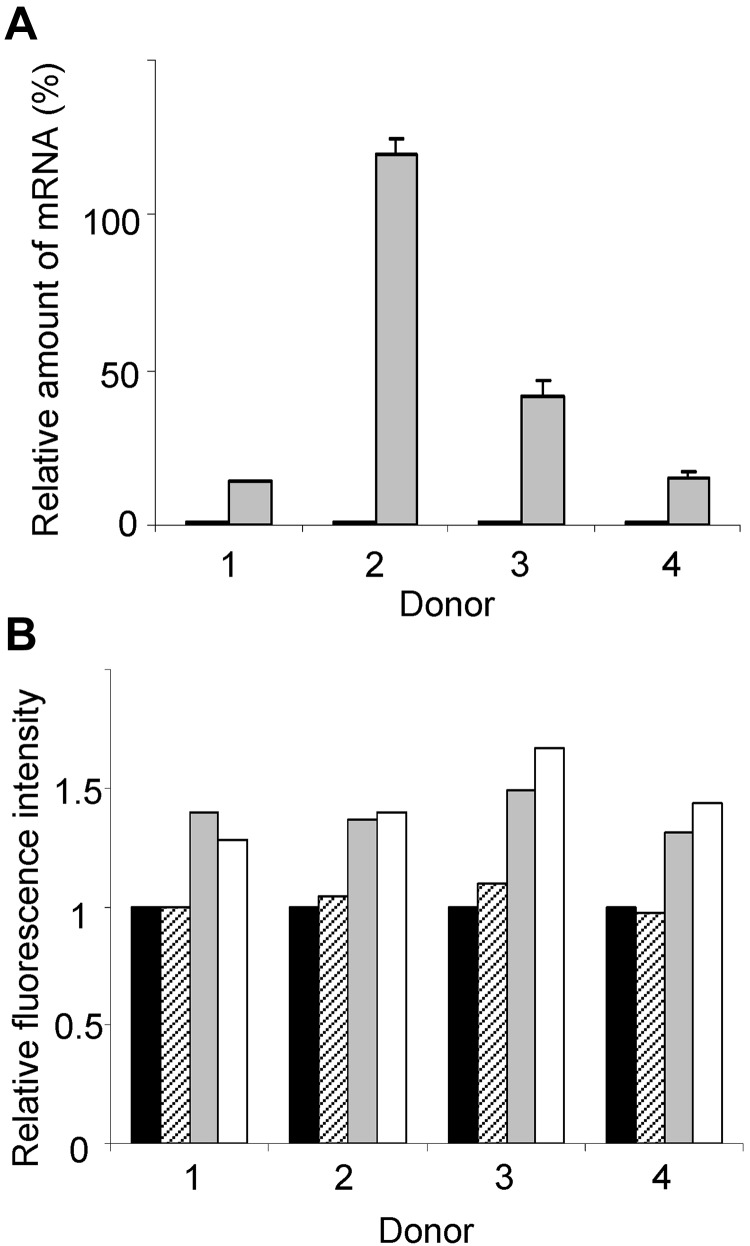
Figure 6.
Elevated KLF13 expression correlates with increased RANTES expression in resting T lymphocytes. (A) PBMCs from 4 donors were treated for 16 hours with (gray) or without (black) CMA (1 μg/mL), and RANTES mRNA levels were determined by real-time quantitative PCR. (B) PBMCs from 4 donors were treated with CMA for 16 hours, and RANTES expression was determined by intracellular staining and flow cytometry. Mean fluorescence is expressed relative to expression in cells cultured in medium alone (black bar). RANTES expression in CD56+ NK cells (hatched bars), CD56+CD3+ NK T cells (gray bars), and CD3+ T cells (open bars) is shown. Data are mean ± SD.

