
Figure 5. The Abl biosensor peptide is present and phosphorylated in both the cytoplasmic and nuclear fractions of Abl-WT-EGFP cells.
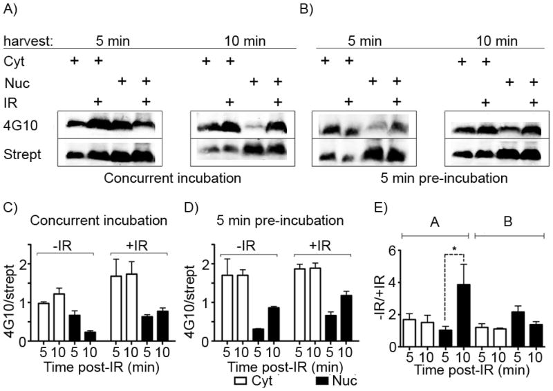
By isolating the cytoplasmic and nuclear material from cells treated with the Abl biosensor within 5 min after IR, we simultaneously examined subcellular localization and the earliest practical timepoint for post-IR sample processing. Western blotting was performed for biotinylated (Strept) and phosphorylated (4G10) peptide from 50 μg/lane of each subcellular fraction from cells incubated with the biosensor peptide A) concurrently with IR treatment or B) pre-treated for 5 min prior to IR. A) In cells expressing Abl-WT-EGFP, the biosensor peptide is observed in both the cytoplasmic (e.g. lanes 1 and 2) and nuclear (e.g. lanes 3 and 4) fractions within 5 min of incubation with and without IR. C) and D) Integrated band intensity data for 4G10 and streptavidin signals over three independent replicate experiments were calculated and the phosphorylated signal (4G10) was divided by the total’ marker signal (streptavidin) and plotted. E) These were further normalized to their change in relative intensity following IR). Y-axes are labelled as 4G10/strept or +IR/−IR, meaning the increase in the 4G10/strept signal for samples treated with IR vs non-IR controls. Error bars represent SEM, and statistical significance (p<0.05) is indicated using *.