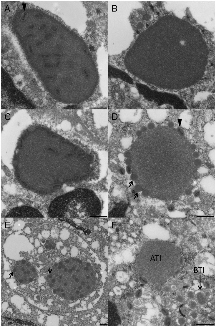
Figure 5. Electron microscopic examination of the inclusion bodies.
Three types of ATIs were observed; inclusions containing virions throughout (Fig. 5A), inclusions without virions (Fig. 5B), and inclusions with virions at the periphery (Fig. 5C). The ATIs examined had varying morphologies that included both non-condensed and mature virions inside and/or around the periphery of the inclusions (Fig. 5D, E). B-type inclusions (BTIs) were also observed (Fig. 5F). The arrow head in figure 5A, and 5D, shows a mature volepox virion; the arrows in figure 5D, and 5E, show immature or non-condensed virions.

