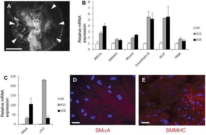
Figure 1. Development of SMCs in human embryoid bodies.
(A) Low power dark field image of day 28 human embryoid body grown in 20% FBS showing outgrowth of cells (white arrowheads) from the central embryoid body mass. (B and C) Expression of SMC specific genes in embryoid bodies by real time RT-PCR at days 0, 15 and 28 is normalised by three housekeeping genes (GAPDH, UBC, 18S) and then presented relative to undifferentiated human ESCs. RT-PCR data represent means from three independent experiments. Bars represent s.e.m. Cells that stain for SMαA (D) and SMMHC (E) clearly seen at day 28 within embryoid bodies by immunofluorescence. Nuclei counterstained (blue) with DAPI. Bar in A = 1000 µm, bars in D and E = 20 µm.

