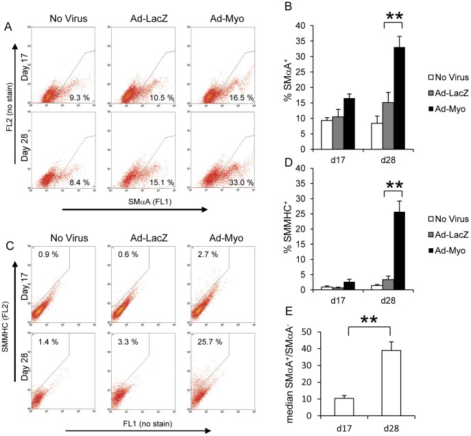
Figure 3. Myocardin overexpression increases number of SMC-like cells.
Embryoid bodies were enzymatically dispersed into single cells at day 17 or day 28 and flow cytometric assessment for SMC markers was carried out. Groups that had been treated with no virus, Ad-LacZ or Ad-Myo from day 10 onwards were used to quantify the proportion of SMαA+ cells (A & B) and SMMHC+ cells (C & D). Both FL1 and FL2 channels were measured for all samples to distinguish specific signal for SMαA (FL1 in A) and SMMHC (FL2 in B) due to the high levels of autofluorescence in embryoid body-derived cells. In the no virus group, SMαA staining was quantified as median SMαA+ signal/median SMαA− signal at both day 17 and day 28 (E). Data presented in A and C are representative flow cytometric plots from a single study with the means from three independent experiments specified in the gated regions and as bar charts ± s.e.m. (B, D & E). **p<0.01.

