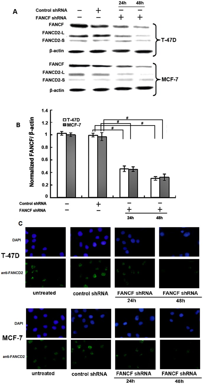
Figure 1. Inhibition of FANCF levels, FANCD2 ubiquitination and foci formation by FANCF shRNA in breast cancer cell lines.
MCF-7 and T-47D cells were transfected with FANCF shRNA and control shRNA(scrambled shRNA) for 24 h and 48 h, then protein was extracted for western blotting with anti-FANCF and anti-FANCD2 antibodies (FANCD2-L = mono-ubiquitinated; FANCD2-S = nonubiquit- inated). β-actin was simultaneously immunodetected to verify equal loading of cell lysates. (A) Representative FANCF and FANCD2 blots. Three independent experiments were performed. (B) Densitometric analysis was done for FANCF expression. Results were normalized to β-actin values. Graphs show means ± S.D. of three independent experiments. P values, # P<0.05. (C) FANCD2 foci formation was detected by immunofluorescence. Representative images are shown.

