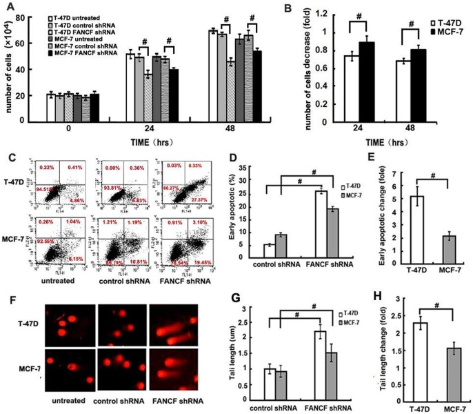
Figure 2. FANCF shRNA inhibits cell proliferation, increases apoptosis in T-47D and MCF-7 cells.
(A) Number of viable cells was determined using a hemacytometer after staining dead cells with Trypan Blue. (B) Quantitative analysis of the fold decrease of total cell number in MCF-7 cells and T-47D cells treated with FANCF shRNA compared with control shRNA. (C) Apoptosis of cells were measured using FACScan after staining with FITC-annexin V and PI. Cells in the lower right-hand quadrant are early apoptotic cells with exposed phosphatidylserine (FITC-annexin V-positive), but intact membrane (PI-negative). (D) The quantification of apoptosis in the indicated cell lines. (F) Single-cell gel electrophoresis (comet assay) showed detectable comet tails when visualized under a fluorescent microscope, indicative of DNA damage. (G) The quantification of DNA fragmentation in the indicated cell lines (control shRNA treated cells was defined as 1.0). (E) and (H) are the quantitative analysis of the fold increase of early apoptotic or tail length in FANCF-silenced cells compared with the controls. P values, # P<0.05.

