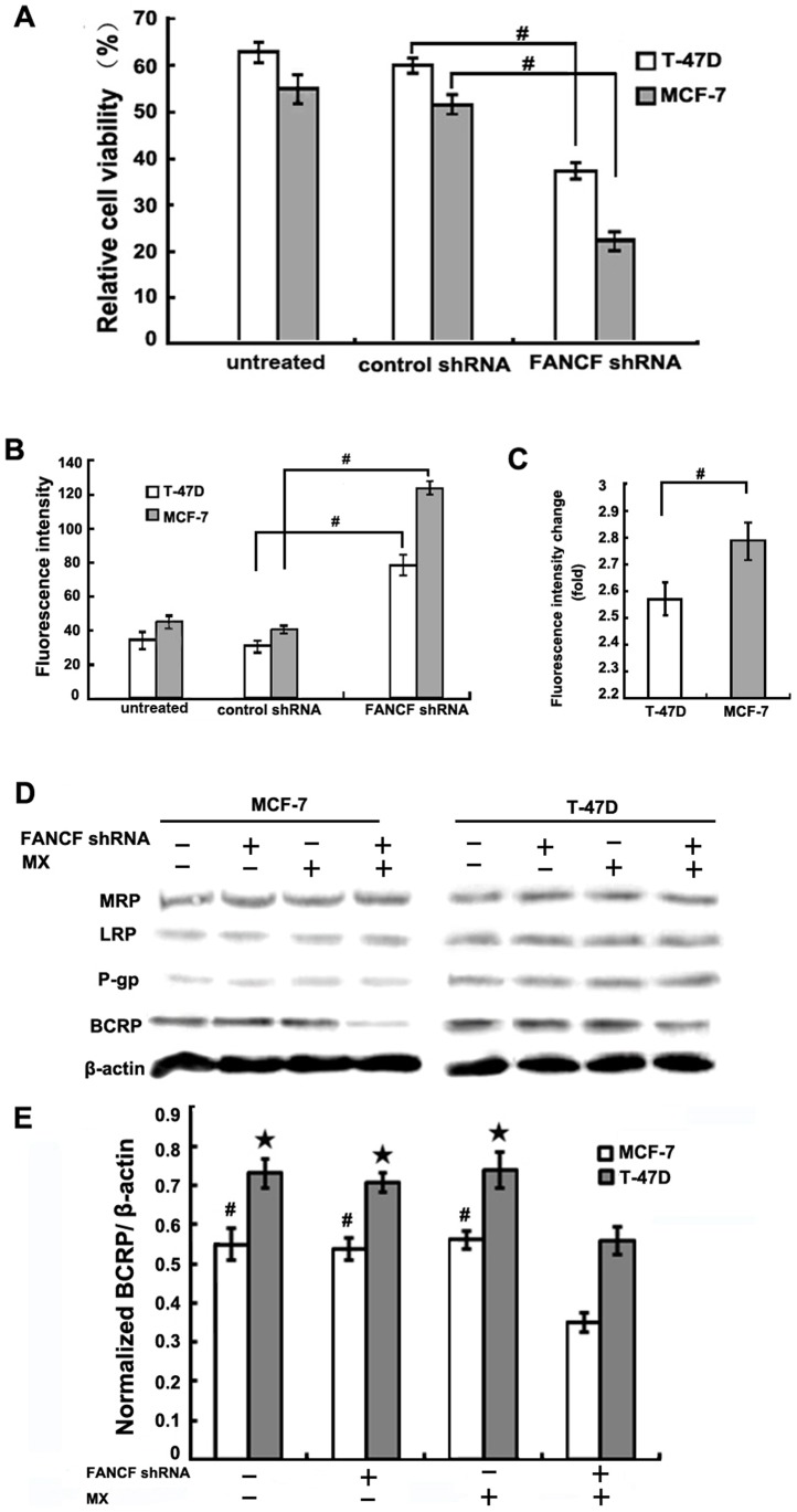
Figure 3. FANCF knockdown sensitizes breast cancer cells to MX.
(A) T-47D and MCF-7 cells were transfected with FANCF shRNA or control shRNA for 48 h, and then treated with 10 uM MX for 24 h. The cell viability was determined by the MTT assay. The percentage of viable cells was determined by the ratio of viable cells treated with MX to that with no MX treatment. (B) Median fluorescene intensity was measured indicating the relative amount of MX accumulation. (C) Quantitative analysis of the fold increase of fluorescence intensity in FANCF-silenced MCF-7 and T-47D cells compared with the controls. P values, # P<0.05. (D) MRP/LRP/P-gp/ BCRP protein expression was detected by western blot assay. (E) Densitometric analysis was done for BCRP expression. P values, # P<0.05 versus transfected with FANCF shRNA and MX in MCF-7 cells,★P<0.05 versus transfected with FANCF shRNA and MX in T-47D cells.

