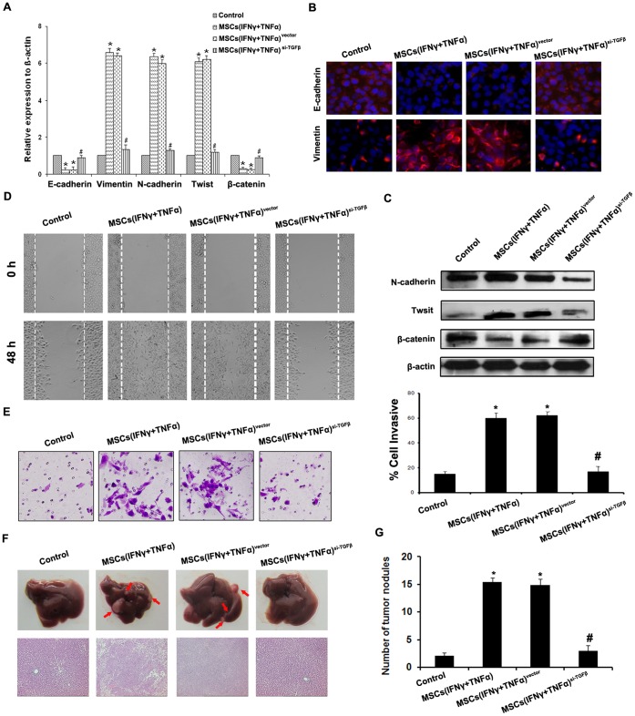
Figure 3. TGF-β depletion in MSCs reverses the promotive effect on metastasis and EMT of HCC cells induced by MSCs in inflammation microenvironment.
(A) Expression of EMT genes was detected by qPCR (normalized to β-actin); (B) E-cadherin and Vimentin expression was performed by immunofluorescent staining, nuclei were counterstained with DAPI (×200). (C) Western blot was used to detect the expression of N-cadherin, Twist and β-catenin, SMMC-7721 cells co-cultured with MSCssi-TGFβ stimulated by both IFNγ and TNFα did not present EMT; (D) The wound healing assay was employed to determine the migration of SMMC-7721 cells (×200); (E) Invasiveness of SMMC-7721 cells was determined using Transwell assay; (F) The metastatic liver nodules in nude mice by splenic-vein injection of SMMC-7721 cells. The arrows indicate the metastatic tumor on the surface of the liver (upper). H&E staining was performed on serial sections of metastatic tumors and normal liver (bottom, ×100); (G) The number of nodules were quantified on livers of nude mice (n = 10 per group) 6 weeks after splenic vein injection of SMMC-7721 cells. (*P<0.05 versus Control group; #P<0.05 versus MSCs(IFNγ+TNFα) and MSCs(IFNγ+TNFα)vector; ×200).

