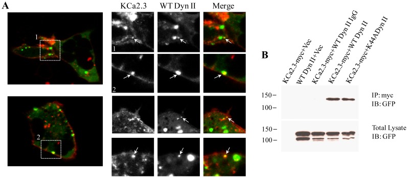
Figure 3. Endocytosis of KCa2.3 is dependent upon dynamin.
A. GFP-tagged WT dynamin II and BLAP-tagged KCa2.3 were expressed in HEK293 cells. KCa2.3 was labeled at the plasma membrane with streptavidin-Alexa555 for 10 min at 4°C after which the cells were immediately imaged by live-cell confocal microscopy at 37°C (see Methods). Left Panels show individual images from two separate cells during the time course of the experiment. Right Panels show cropped images from these two cells (labeled 1 and 2) as well as cropped images from 2 additional cells. Arrows denote co-localization of KCa2.3 and WT dynamin II. B. Co-IP of myc-tagged KCa2.3 with GFP-tagged dynamin II. KCa2.3 was immunoprecipitated using an anti-myc Ab (lanes 1, 2, 4, 5) or an anti-V5 Ab as IgG control (lane 3) and subsequently IB using an anti-GFP Ab for dynamin II. WT and K44A dynamin II were detected by IB in lanes 4 and 5, respectively, confirming an association between KCa2.3 and dynamin (Top Panel). Bottom Panel confirms expression of GFP-tagged dynamin in total lysate by IB (5 µg total protein loaded per lane). Data are representative of 3 experiments.

