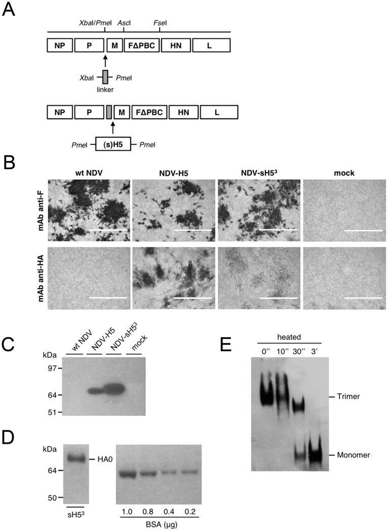
Figure 1. Construction of recombinant NDV, expression and purification of H5 protein.
A) Construction of pFL-HertsΔPBC expressing influenza H5N1 HA proteins. The sequence of the full-lenght cDNA of Herts/33, which codes for the multibasic cleavage site of the F protein was mutated into a monobasic cleavage site-encoding sequence (FΔPBC) resulting in pFL-HertsΔPBC. The HA gene of H5N1 A/Vietnam/1194/04, encoding either the full length or the soluble trimeric form, was cloned between the PmeI sites created in the intergenic region of the P and M genes of the pFL-HertsΔPBC vector following insertion of a linker containing NDV transcription termination and start signals. NP, nucleoprotein; P, phosphoprotein; M, membrane protein; F, fusion protein; HN, hemagglutinin-neuraminidase; L, large polymerase. B) Expression of HA protein by recombinant NDV in QM5 cells. Cells were infected with wt NDV (Herts/33), NDV-H5, NDV-sH53 or were mock-infected and analyzed by immunocytochemistry using mAb specific for NDV F or influenza virus H5. Scale bars represent 400 μm. C) Western blot analysis with an anti-H5 mAb following SDS-PAGE of combined lysate of QM5 cells and the cell culture supernatant (20 μl/T75 flask). The MW of the marker proteins is indicated on the left. D) The sH53 protein was purified from the supernatant of NDV-sH53-infected QM5 cells and subjected to SDS-PAGE (∼ 1 μg/15 μl), after which proteins were stained with Blue stain reagent. A series of BSA dilutions were run in parallel. E) Blue native PAGE analysis of sH53 purified from the cell culture supernatant of NDV-sH53-infected QM5 cells (∼ 1.7 μg/20 μl). The position in the gel of the trimeric and monomeric form of the H5 protein is indicated. Prior to gel electrophoresis some samples were heated at 95°C for the indicated time periods.

