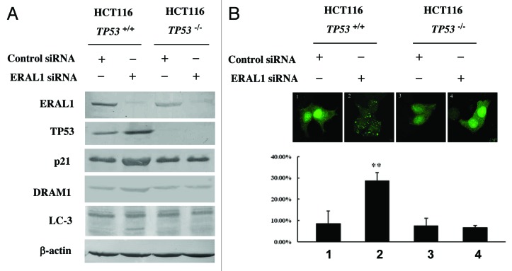
Figure 5. TP53 is essential for the autophagy induction by ERAL1 knockdown. (A) HCT-116 TP53+/+ and HCT-116 TP53−/− cells were transfected with ERAL1 siRNA. At 72 h post-siRNA transfection, cells were subjected to western blotting to detect the levels of the indicated proteins. (B) HCT-116 TP53+/+ and HCT-116 TP53−/− cells were transfected with GFP-LC3 plasmid and then treated with control siRNA or ERAL1 siRNA, respectively. At 72 h post-siRNA transfection, GFP-LC3 puncta formation in the cells was detected by confocal microscopy. The percentage of GFP-LC3 puncta-positive cells was quantified as described under Materials and Methods. Representative data were from three independent experiments. The p value derived from Student’s t-test is **p < 0.001.

An official website of the United States government
Here's how you know
Official websites use .gov
A
.gov website belongs to an official
government organization in the United States.
Secure .gov websites use HTTPS
A lock (
) or https:// means you've safely
connected to the .gov website. Share sensitive
information only on official, secure websites.
