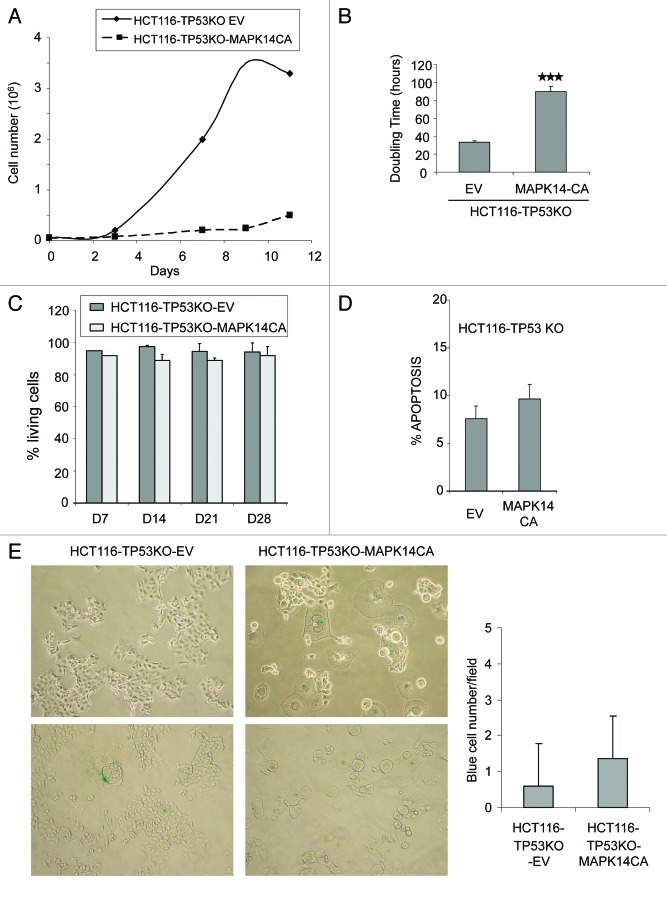
Figure 2. Overexpression of constitutively active MAPK14 inhibits cell growth in HCT116-TP53KO cells. (A) Growth curve of HCT116-TP53KO-EV and HCT116-TP53KO-MAPK-CA cells cultured in complete medium (10% FCS). (B) Doubling time histograms of HCT116-TP53KO-EV and HCT116-TP53KO-MAPK14-CA determined at day 14 after retroviral transduction of MAPK14-CA or EV. (C) Percentage of living cells in HCT116-TP53KO-EV and HCT116-TP53KO-MAPK14-CA cells at day (d) 7, 14, 21 and 28 after retroviral transduction. The number of viable cells was determined by counting the number of cells not stained by the Trypan blue dye. Data are representative of three independent experiments. (D) Quantification of apoptosis in HCT116-TP53KO-EV and HCT116-TP53KO-MAPK14-CA cells at day 14 after retroviral transduction of MAPK14-CA or pMSCV empty vector (EV). Apoptosis was determined by quantifying the number of 7AAD-negative and Annexin V-FLUOS-positive cells with a FACScan flow cytometer. (E) Senescence-associated β-galactosidase staining in HCT116-TP53KO-EV and HCT116-TP53KO-MAPK14-CA cells at day 14 after retroviral transduction of MAPK14-CA or EV. The number of β-galactosidase-positive (blue) cells was counted in 20 fields for each cell type.

An official website of the United States government
Here's how you know
Official websites use .gov
A
.gov website belongs to an official
government organization in the United States.
Secure .gov websites use HTTPS
A lock (
) or https:// means you've safely
connected to the .gov website. Share sensitive
information only on official, secure websites.
