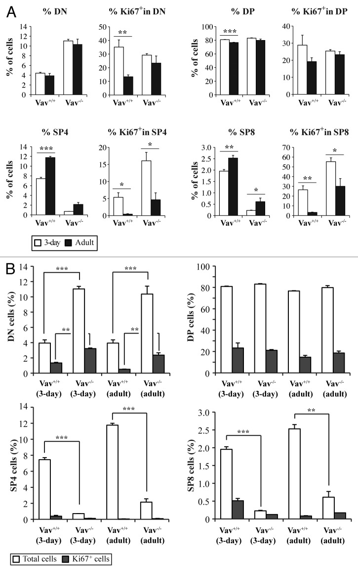
Figure 5. Cell cycle entry of different thymocyte populations. Thymocytes were stained with the corresponding antibodies to identify CD4-/CD8- (DN), CD4+/CD8+ (DP), CD4+/CD8- (SP4) and CD4-/CD8+ (SP8) cells and with an anti-Ki67 antibody to analyze cell cycle entry. A) The total percentages of cells and the percentages of Ki67+ cells in the different populations are shown in 3-d old (white bars) and adult (black bars) mice of both genotypes . B) Comparative analysis of Ki67 expression in the different populations between Vav1−/− and wild-type mice. Data represent the mean percentages ± SEM of three independent experiments with n = 3 mice per group. Data were evaluated using Student’s t-test: * p < 0.05; **p < 0.01; ***p < 0.001.

An official website of the United States government
Here's how you know
Official websites use .gov
A
.gov website belongs to an official
government organization in the United States.
Secure .gov websites use HTTPS
A lock (
) or https:// means you've safely
connected to the .gov website. Share sensitive
information only on official, secure websites.
