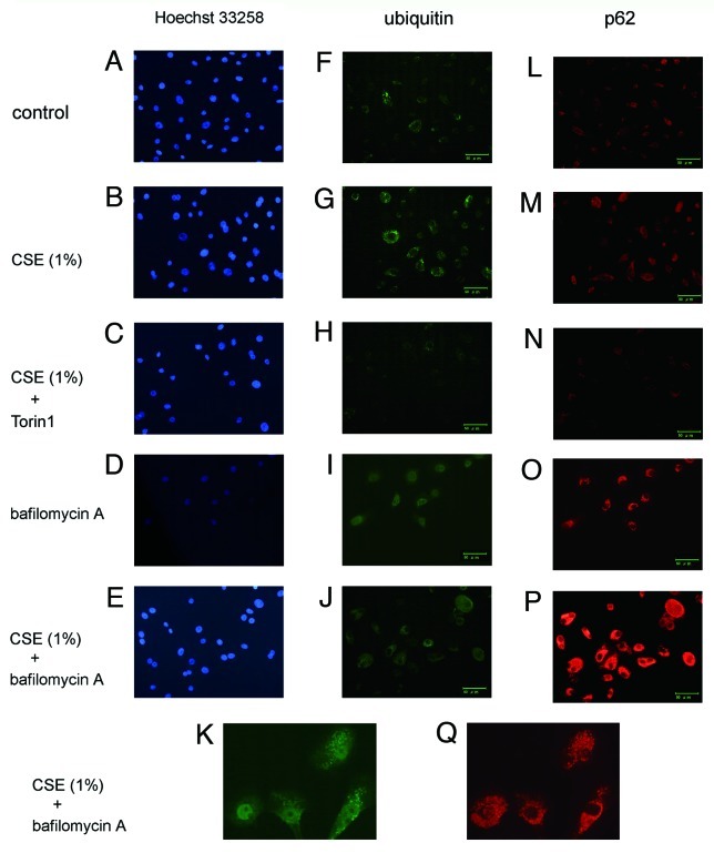
Figure 5. Immunofluorescence staining of ubiquitin and p62 in HBEC. Photomicrographs of nuclear staining with Hoechst 33258 (panels A to E), immunofluorescence staining with anti-ubiquitin (panels F to K), and anti-p62 (panels L to Q). HBEC were control treated (A, F, L) or CSE (1.0%) treated (B, C, E, G, H, J, K, M, N, P, and Q), in the presence of Torin1 (250 nM)(C, H, N), or bafilomycin A (200 nM) (D, E, I to K, O to Q). All photomicrographs are taken at the same magnification. Original magnification is × 200. Bar = 50 μm. K and Q are high magnification view.

An official website of the United States government
Here's how you know
Official websites use .gov
A
.gov website belongs to an official
government organization in the United States.
Secure .gov websites use HTTPS
A lock (
) or https:// means you've safely
connected to the .gov website. Share sensitive
information only on official, secure websites.
