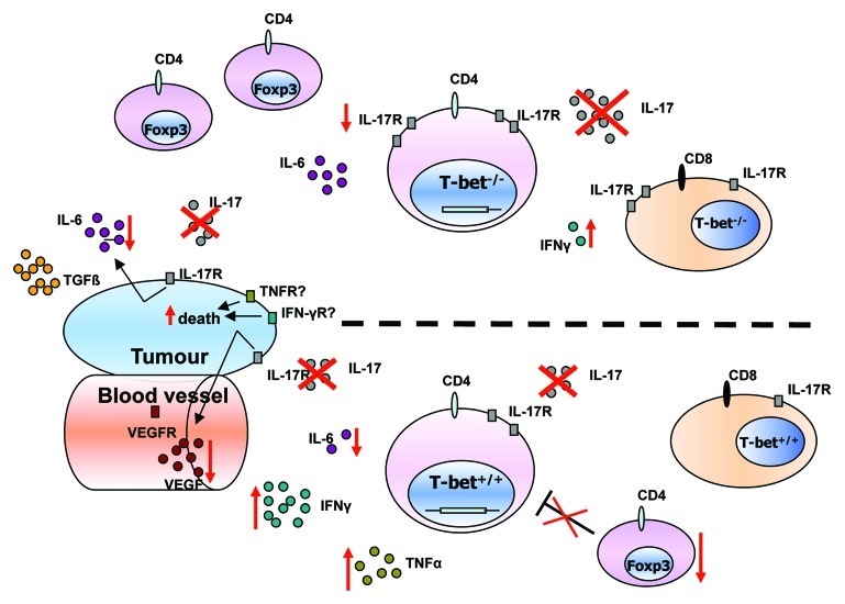
Figure 1. Effect of anti-IL-17A antibody treatment in the absence and presence of T-bet in experimental non-small-cell lung cancer. Immunological cytokine milieu in T-bet−/− (upper part) and wild-type (lower part) tumor bearing mice. The number of circles for each cytokine or rectangles for receptors, reflect their relative amount in T-bet−/− and wild-type mice. Red crosses represent the blockade of IL-17A. Red arrows indicate an upregulation or downregulation of cytokines, receptors or number of cells after IL-17A blockade. T-bet−/− mice show higher IL-17A, IL-6 and IL-17R expression and exhibit more T-regulatory cells whereas IFNγ levels are decreased. After IL-17A blockade there is an upregulation of IFNγ and TNFα secretion by CD4+ T-cells and a downregulation of IL-6 and Foxp3+ T cells in the lung of wild-type mice bearing tumor. In T-bet−/− mice IL-17A blockade led to a downregulation of IL-17R expression on CD4+ T-cells and an upregulation of IFNγ produced by CD8+ T-cells in the lung of mice bearing tumor.

An official website of the United States government
Here's how you know
Official websites use .gov
A
.gov website belongs to an official
government organization in the United States.
Secure .gov websites use HTTPS
A lock (
) or https:// means you've safely
connected to the .gov website. Share sensitive
information only on official, secure websites.
