
Figure 1. NFIA expression during oligodendrocyte development.
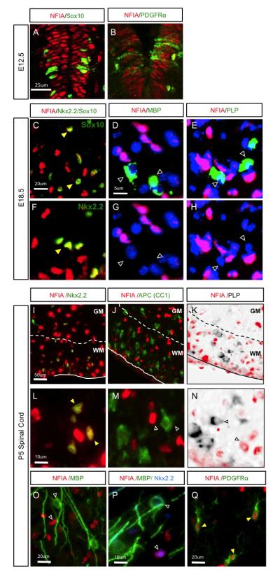
(A-B) NFIA is co-expressed with Sox10 and PDGFRa in the spinal cord at E12.5. (C-H) NFIA is co-expressed with OLP markers Sox10 and Nkx2.2 at E18.5 (C,F), but not markers of mature OLs MBP (D,G) or PLP mRNA (E,H). Images in C and F are from distinct areas and samples; Image sets in (D,G) and (E,H) are from the same image. (I Q) Analysis of NFIA expression in the post-natal spinal cord. NFIA is co-expressed with OLP marker Nkx2.2 (I,L,P), but not differentiation marker APC (J,M) or mature markers PLP (K,N) and MBP (O,P). In I-K, the dashed line denotes the border between the white matter (WM) and the grey matter (GM). Filled-yellow arrowheads indicate colocalization. Unfilled arrowheads indicate lack of colocalization.