
Figure 2. Expression of NFIA in oligodendrocyte progenitors in human HIE and MS.
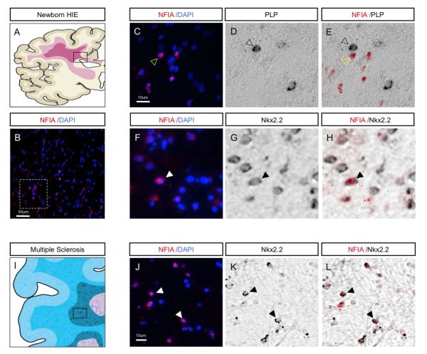
(A) Schematic illustrating white matter damage in pediatric HIE. Boxed area represents region of gliotic sub-cortical white matter and origins of the tissues used for analysis. (B) Expression of NFIA in human HIE; dashed box is region shown at higher magnification in C. (C-H) NFIA is not co-expressed with PLP-expressing cells (C-E), but is co-expressed with Nkx2.2-expressing cells (F-H), indicating expression in OLPs within areas of affected white matter. (I) Schematic demonstrating affected areas of white matter in human MS. Boxed area represents active lesion site and region used for analysis. (J-L) NFIA is co-expressed with Nkx2.2 within active MS lesions, indicating expression in OLPs. Filled arrowheads indicate colocalization. Unfilled arrowheads indicate lack of colocalization.