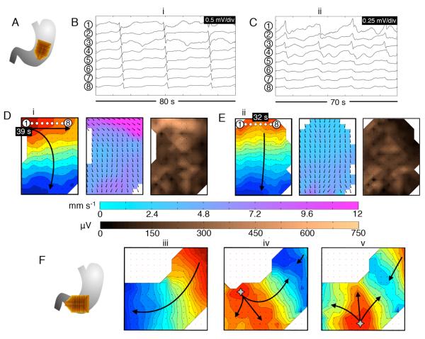
Figure 5.
Abnormal slow-wave initiation at low frequency. A. Position diagram relating to (B-E); diabetic gastroparesis (ID#1). B,D. Stable ectopic pacemaking originated near the lesser curvature of the mid-corpus, at a slightly lower than normal frequency (2.4 ± 0.05 SD c/min). Isochronal (intervals = 1s), velocity and amplitude maps are shown for typical wave (i) (see also animation Figure5i.wmv). C,E. The ectopic pacemaker was transient, being followed by activity of normal pattern and frequency (3.0 ± 0.05 SD c/min). Maps are shown for representative wave (ii) (see also animation Figure5ii.wmv). During ectopic pacemaking, circumferential slow-wave propagation occurred, and was faster than normal longitudinal conduction (10.7 ± 6.2 SD vs 3.3 ± 1.5 SD mm s−1; P<0.001), with higher amplitudes (270 ± 155 SD vs 80 ± 40 SD μV; P<0.001). The time stamps are marked relative to the animations. F. A further example of propagation patterns during bradygastric activity, mapped at the corpus-antrum border, in idiopathic gastroparesis (ID#9). Normal activity (frequency 2.9 ± 0.1 SD c/min); e.g. wave (iii), was followed by a period of unstable ectopic focal events at a lower than normal frequency (2.2 ± 0.5 SD); waves (iv, v), which caused retrograde-propagating wavefronts that collided with proximal activities.

