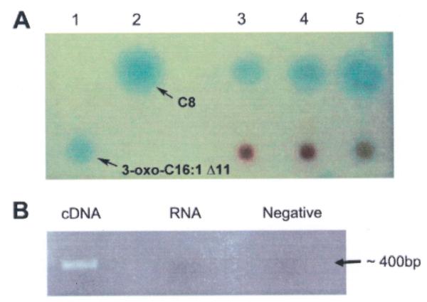
Figure 4. Detection of ssaI gene expression and AHLs in sponge tissue.

A) Reverse phase C18 thin layer chromatography plates overlayed with A. tumefaciens AHL reporter for detecting AHLs from sponge tissues. Lane 1: 3-oxo-C16:1 Δ11, lane 2: C8-HSL, lanes 3-5: M. laxissima individuals 1-3. B) RT-PCR detection of expression of ssaI gene in sponge tissue. The PCR amplicon is about 400 bp. The first lane used cDNA as template, the second lane used RNA as template to test for DNA contamination (RNA control) and the last lane was a no template negative control.
