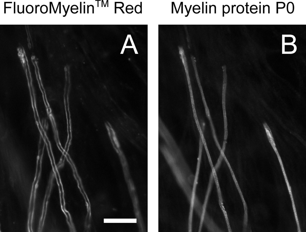
Fig. 4. FluoroMyelin™ Red stains all myelin sheaths.
Myelinating cultures were incubated for 2 hours in observation medium containing FluoroMyelin™ Red (1:300 dilution) after 34 days in culture. After staining we imaged random fields in fresh observation medium using a 40× objective magnification and then we fixed the cultures and imaged the same fields again after processing for immunofluorescence with an antibody to the myelin protein P0 (see Methods). A. FluoroMyelin™ Red fluorescence in living culture. B. Myelin immunofluorescence after fixation. Note that there was some movement and shrinkage of the axons and myelin sheaths during fixation and subsequent processing, but nevertheless it is clear that there is a close correspondence between the FluoroMyelin™ Red fluorescence in the living cultures and the myelin fluorescence in the fixed cultures. Scale bar=10 µm.

